


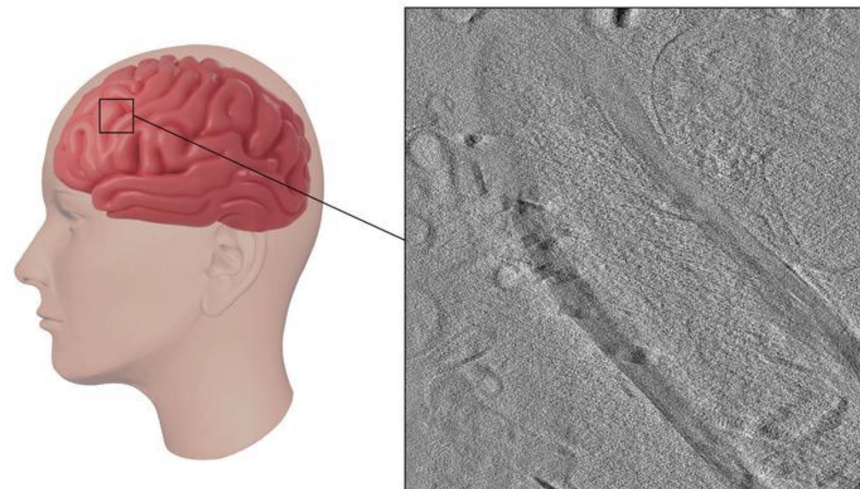
There are amazing advances in the technology used on the human brain. A new technique uses ion beams to view structures inside human brain cells. Ultimately, this may help us to understand brain diseases, including Alzheimer’s or multiple sclerosis.
Scientists today use electron microscopy, which involves adding chemicals and physically cutting the tissue. The challenge is that the approach can change how the cells and structures look and limit resolution. Another method, cryo-electron tomography, provides clearer images of the brain’s smallest parts in a more native state, but it requires freezing samples to cryogenic temperatures, and if done incorrectly, ice crystals can form.
Research at the University of Pennsylvania shows a new technique to study the human brain’s ultrastructure. The technique will be presented at the 68th Biophysical Society Annual Meeting in Philadelphia, Pennsylvania.
The team obtained brain tissue from autopsies, flash-froze it directly on special grids with liquid ethane, and used a xenon plasma-focused ion beam (FIB) to cut thin slices for imaging. This method allowed them to look at the brain tissue in its near-natural state, eliminating cutting with a knife or adding chemicals. The new method enables easy and rapid freezing of much thicker samples. Prior methods were limited to 10 microns; the new method allows for 250 microns thick without ice crystals and is substantially faster.
The approach enables the observation of intact structures within cells, such as tau fibrils, a hallmark of Alzheimer’s disease, and cellular components that try to break down these fibrils. Researchers also visualized and measured myelin, a sheath critical for nerve function that breaks down in certain diseases, such as multiple sclerosis.