
To prevent the drying and deforming of biological samples, a nanosheet made of organic polymers has been developed, thus enabling high-quality imaging under microscopes.
Be it cosmology or biology, the advancement of science largely relies on the advancement of measuring instruments and methodology. In the past couple of decades, scientists’ passion to see the invisible has vastly improved microscopes and other equipment resulting in high-resolution images, three-dimensional images, and longer recording times of biological samples.
However, current setups do not prevent them from drying and deforming during observations, resulting in blurred images.
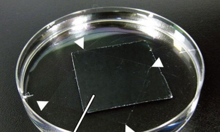
Scientists from Hokkaido University and Tokai University in Japan have developed a nanosheet made of a fluorine-containing polymer known as CYTOP and investigated its effectiveness in producing clear images when wrapped around biological samples. CYTOP is a commercially available chemical agent developed by Asahi Glass.
The researchers successfully produced a nanosheet of CYTOP by spreading its solution on a layer of Poly-Vinyl-Alcohol (PVA). The nano-thin layer of CYTOP was easily detached from the PVA layer by soaking them in water. The thickness of the nanosheet could be adjusted by changing the concentration of its solution. The nanosheet retained CYTOP’s high water repellent properties which should help retain a sample’s water content when used as wrapper.
The team then tested the nanosheet’s wrapping properties by using alginate hydrogel to simulate biological samples such as tissue. They found that the gel retained 60% of its water content after 24 hours when wrapped whereas the unwrapped gel became totally dehydrated after about 10 hours. Experimenting with various thicknesses, they found that a 133nm-thick sheet provides sufficient water retention and surface adhesion which is necessary for fixing samples to the cover slip.
They next applied an actual biological sample: 1mm thick brain slices from mice. Without a CYTOP wrap, evaporation of the embedded water caused local, non-uniform sample shrinkage, leading to blurred images. By wrapping the brain slices in a CYTOP nanosheet, however, images with a high spatial resolution could be obtained from scanning a large area (more than 750×750μm) over a long time (about 2 hours). Similar results were obtained even when the sample was embedded in agarose gel – a common technique used for mounting biological tissues that could disturb the clarity of taransparentized samples — without disturbing the clarity.
“The wrapping technique is still at an early stage, but it establishes and verifies the superiority of nanosheet wrapping for tissue imaging,” said the researchers of the paper published in Advanced Materials.